Respiratory Physiology FAQs - selected Q/A from/to students
2010
Hi Dr. Mayrovitz, I had a question about your P-V compliance graph on page 14:
I understand that the X-axis is showing recoil pressures for the chest,lung etc.
But if these recoil pressures are mathematically equivalent to Translung,
transwall etc. why is there is discrepancy in how the pressures functionally
change with lung volume. Specifically, my question is if Transwall (Tw)
is equal to intrapleural (Ppl) as you derived on page 13 (top) why is the
Tw getting less negative with increasing lung volume? (Tw) would not be
equivalent to (Ppl) on this graph, why is this?
Thank you for your response (many people asking ).
DR. M replies
The graphic at the bottom of page 14 shows how chest wall and lung recoil pressures
(x-axis) depend on lung volume (y-axis). Recoil pressures can have +, - or 0
values depending on their direction of action. Recoil pressures in the
inward direction (chest or lung wants to get smaller) are defined as positive and
outward recoil tendencies (chest only) are defined as negative. Although transwall
pressure (Ptw) is numerically equal to the intraplerual pressure (Ppl) as noted on
page 13, the DIRECTION of the chest recoil pressure depends on lung VOLUME not
necessarily on Ptw. The reason for this is that the recoil is dependent on the
displacement from the equilibrium (zero stress) state. This concept is illustrated
by the top left figure of page 14. Thus it would be possible to have differing
lung or chest conditions in which the same value of Ptw was associated with
different lung volumes and thereby different recoil pressures. See also the
information provided to a similar question
Dear Dr. Mayrovitz,
The BRS book (I know, I am sorry), says that Airway obstruction, or shunt
results in a low V/Q ratio, whereas pulmonary embolus results in an infinite
V/Q ratio, and that an infinite V/Q ratio represents dead space. Is this not
correct? I have been trying to sort this out all day, and I just seem to
remember you explaining it differently in class. Thank you again for your
time.
Dr. M replies
If Q is zero indeed V/Q is infinite and the region for which this is true is indeed dead
space. But this only applies to the region that has the zero flow. In the example
presented in lecture the effect of the flow blockage was shown to cause an increase in
flow in the other lung (blocked flow was diverted) causing the V/Q ratio of the total
lung to decrease thereby lowering the net PO2 in the blood exiting the lung.
Dear Dr. Mayrovitz
Ok. Thank you very much for your answers. So, is it possible for an
embolus to result in V/q increase or decrease i.e., v/q = zero) or
a Dead space (V/q=infinity) pattern? What about an airway block? Could
it result in either a V/q increase or decrease, or only a decrease?
I think my confusion is coming in part from the pictures at the bottom
of page 37. It shows that an airway obstruction causes reduced V/Q
ratio.
Is a V/Q ratio of zero called a shunt? The BRS book says that a V/Q
ratio of zero is called a shunt, whereas a V/q of infinite is called
deadspace.
Furthemore, it says that shunts are caused by airway obstruction, and Deadspace is caused by pulmonary embolism.
This is not entirely correct according to what we have learned in class?
Thank you
Dr. M Replies
Correct - their distinctions can be confusing - perhaps even inaccurate.
Dead space in this context refers to the functional aspect of the alveoli.
Alveoli without ventilation but adequate flow represent dead space since these alveoli do
not contribute to alveoli-blood gas exchange. V/Q = 0
Alveoli with ventilation but no blood flow also represent dead space since any
ventilation of these alveoli is wasted ventilation since it does not contribute to
alveoli-blood gas exchange. In this case the V/Q ratio for the involved alveoli and their
associated blood capillaries is infinite because the flow (perfusion) is zero.
Since the flow is zero this blood does not directly effect the net PaO2 of the pulmonary
vein blood. However, since the blood that would have gone to the blocked region is
diverted to other regions the V/Q value of these other regions becomes
reduced thereby causing the PaO2 to be less than normal. The limit case is when an
entire lung is blocked by the embolis.
Dear Dr. Mayrovitz,
I need some help understanding this concept..hopefully you can help me out..
what is the effect of intrapleural pressure on blood flow..specifically which
way does it push or pull..
and in general can you describe the force that intrapleural pressure exerts in
the lung...for example i know that recoil pressure acts to squeeze lung
alveoli...i just have tough time understanding this intrapleural pressure and
its regional effects on blood flow (bottom of page 36)
..is the intrapleural pressure more negative int the base of the lung?
Dr. M replies:
Intrapleural pressure (ip) effects blood vessels since it contributes to their surround
pressure and thus to their transmural pressure. If ip decreases then all else the same
the vessel transmural pressure increases causing an increase in vascular diameter and
hence a decrease in vascular resistance and thus a greater blood flow.
In the dependent lung (i.e. standing) the intrapleural pressure due to gravity would be
greater at the base than at the apex. However, gravity also effects the intravascular
pressure in the same direction but with a greater effect since the density of the blood
is greater than the effective density of the ip space. Thus the net effect is have the
vessel transmural pressure at the base be greater than at the apex.
These concepts are illustrated for ip on page 26 of the notes and for the intravascular
pressures on page 33 of the notes.
2009
01/09/2009
Dr. Mayrovitz,
I have a question regarding the section titled "Dynamic Pressure and Flow Changes"
on page 11. Can you explain why the alveolar pressure decreases as air enters the
alveoli, and alveolar pressure increases as air exits the alveoli?
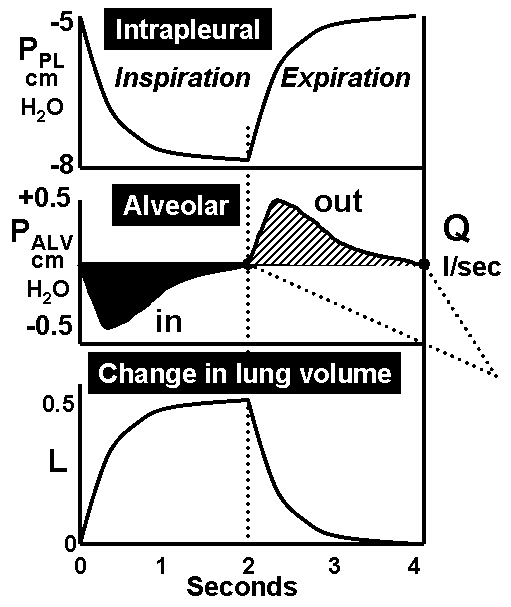 The
adjacent figure is the figure in question. The middle panel
shows the variation in alveolar pressure. The best way to understand
the relationship of this pressure to the air flow (Q) is to recall that
the driving pressure for air flow is the pressure difference between
the atmosphere and the alveoli. When Palv is less than Patm we
have Qin ~ Patm-Palv and when Palv is greater than Patm we have Qout ~
Palv-Patm.
The
adjacent figure is the figure in question. The middle panel
shows the variation in alveolar pressure. The best way to understand
the relationship of this pressure to the air flow (Q) is to recall that
the driving pressure for air flow is the pressure difference between
the atmosphere and the alveoli. When Palv is less than Patm we
have Qin ~ Patm-Palv and when Palv is greater than Patm we have Qout ~
Palv-Patm.
The action of the respiratory muscles acting on the thorax during
inspiration causes the intrapleural pressure (Ppl) to decrease thereby
tending to expand the alveoli since now the alveoli have an increased
transmural pressure. This expansion causes Palv to decrease as
shown in the figure. Since Palv is < Patm the gradient for airflow
is inward. As the alveoli expand their recoil pressure (Prec)
increases since they are being 'stretched' more. The result is
that Palv then begins to turn back toward zero since its value is
determined by the sum of Prec and Ppl. This results in the time coures
of Palv as shown in the figure. But, as long as Palv is <
Patm, airflow is directed inwards. When the inspiratory muscles stop
contracting at the end of inspiration Ppl starts to return
toward its end expiratory value (less negative) and Prec then
starts to decrease as alveolar stretch becomes less. This combo results
in the Palv time course as shown because Palv = Prec + Ppl. As
long as Palv is > Patm, airflow is directed outwards.
To fully analyze the details of the time course of Palv would require a
much more complex story that is not necessary for our purposes.
HNM 01/09/2009
01/09/2009
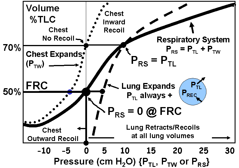
Dr Mayrovitz
On the respiratory compliance P/V relations chart
why does the transwall pressure increase
with chest expansion since
according to the formula TW pressure= pleural-Body surface,
if the thorax expands the transwall pressure should become more negative?
Why does the PRS become more positive with thorax expansion? since
PRS=alveolar pressure, the alveolar pressure should be more negative
upon thorax expansion.
thanks and have a good weekend
 The adjacent figure is the figure in question.
The transwall pressure is in general as you state equal to the
difference between Ppl and Pbs. You are also correct about the
increase (more nagative) of Ppl with increasing volume. Perhaps
the point of confusion arises because it was not made clear that the
x-axis of the graphic is Recoil Pressure
- either of the lung alone, the chest wall alone or in combo. Recoil
pressure is taken as positive when it is in a direction acting inwards
and negative when it is acting outward. Concerning the
chest, as long as the chest wall expansion is less than its zero
stress point then the recoil force is inwards - hence its negative
value. Beyond this point its recoil is inwards hence the positive
value. So, as Ppl becomes more negative as lung volume increases the
chest wall recoil pressure becomes less and less negative , then zero
and then positive. Because the lung recoil is always inward its
pressure is always positive. When the chest and lung recoil pressures
are combined to give the respiratory system recoil pressure the result
is the solid curve. Note that FRC is defined as the condition where the
respiratory recoil pressure is zero.
HNM 01/09/2009
01/09/2009
Dr. Mayrovitz
I was wondering with C rs , why is it in series when the equation is
1/C rs = 1/C L + 1/C w ?
Because it is reciprocal, why is it not in parallel? I thought series summed directly.
Thanks
Although resistances in series sum directly, when compliances are in series they
sum reciprically as 1/Ct = 1/C1 + 1/C2 + .........
The reason has to do with the physical equations that describe the situation.
For those interested in the derivation any standard physics book should be consulted
HNM 01/09/2009
2008
Dr. Mayrovitz,
I had a quick question regarding the Respiratory Gas Partial
Pressures Table on page 17. Does the "Arterial Blood" column
correlate to the Pulmonary Vein and then the 'Venous Blood" column
correlate to the Pulmonary Artery? I was getting confused because the
Pulmonary Artery is carrying systemic blood from RV to the lungs, so
it should be low in oxygen and high in CO2, but those numbers
correlate to the Venous Blood column.
Thank you for your assistance,
Arterial blood = blood exiting pulmonary capillary
Venous blood = blood entering pulmonary capillary
Arterial and venous in the above are referring to systemic
You are correct in you designation/descriptions using the
pulmonary artery and pulmonary vein as to and away from the lung
HNM 01/13/2008
Dr. Mayrovitz,
I am having a bit of trouble with a few things.
1) on page 6, section 9.0 you make a reference to "puffing" where a person with
emphysema might breath through pursed lips to increase the upper airway resistance so
that the lower airways won't close as soon and the person can exhale a greater volume.
How does the increase in upper airway resistance effect the lower airway resistance such
that the lower airways won't close?
2) page 19, under Time Constant and Uneven Ventilation
How does increased resistance lead to a faster filling of the lungs. Is it just that the
pressure of recoil is such that the person is forced to stop inspiration?
Thank you for your assistance.
Sincerely
1) For a given airflow out the higher resistance of the mouth (pursed lips)
causes the pressure in the smaller airways to be larger . This causes an
increase in their transmural pressure that holds them open to a lower volume
than would occur without the pursed lips.
2) Increased R does not lead to faster filling - the figure indicates
a slower filling.
HNM 01/13/2008